Table of Contents
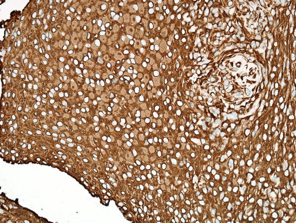
Washington University Experience | NEOPLASMS (MENINGIOMA) | Rhabdoid | 3E Meningioma, atypical w rhabdoid features (Case 3) VIM area A 40X 2.jpg
VIM immunohistochemically stained sections show strong, diffuse immunoreactivity for vimentin in the tumor cells; the eccentric whorls of intermediate filaments are less-intensely stained than the cytoplasm. ---- Ancillary data (not shown): Reactivity for proliferation marker Ki- 67 (MIB-1 antibody) stains a regionally variable proportion of tissue nuclei, of estimated greatest density to be 9.2%; although this area also shows the greatest mitotic density, some of these Ki-67 positive nuclei may be contributed by chronic inflammatory cells. ---- FISH studies are negative for loss/deletion of 1p, 14q, and 9p21 (CDKN2A locus). In sporadic meningiomas, these deletions are more commonly associated with higher grade and less-favorable prognosis. Whether these associations might hold true in the context of pediatric neurofibromatosis type 2 is unclear. Regardless, these negative test results do not provide independent support for more aggressive behavior. ---- Comment: These histomorphological and immunohistochemical features – particularly the presence of four (out of five) atypical histomorphological features (sheeted architecture, hypercellularity, small cell change, and spontaneous necrosis) – support a diagnosis of ‘Atypical meningioma, WHO Grade 2.’since it lacks other anaplastic features, the most appropriate diagnosis for this case is: ‘Atypical meningioma with rhabdoid features, WHO Grade 2.’

