Table of Contents
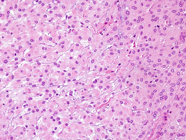
Washington University Experience | NEOPLASMS (MENINGIOMA) | Rhabdoid | 4A5 Meningioma, focal rhabdoid (Case 4) H&E 1 RES Focal R.jpg
Additional tumor image (H&E) Ancillary studies (not shown): A panel of immunohistochemical stains was performed. The vimentin immunostain shows diffuse and strong staining in tumor cells and show pale staining in some of the rhabdoid cells. Ki-67 immunostain shows a variably elevated proliferative index focally up to 7.3%. Progesterone receptor immunostain shows strong positive reactivity in most tumor cells. The overall finding is consistent with meningioma with focal rhabdoid growth and increased proliferation index, WHO grade 1. Although the current tumor does not meet the WHO criteria of a higher grade meningioma, the elevated proliferation index may portend more aggressive behavior.

