Table of Contents
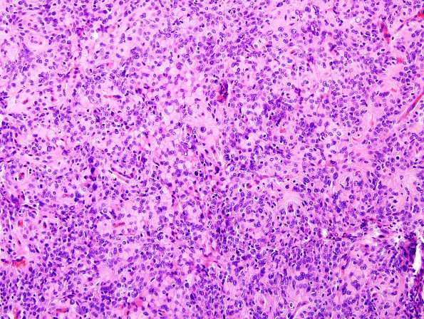
Washington University Experience | NEOPLASMS (MENINGIOMA) | Rhabdoid | 5A1 Meningioma, atypical w rhabd (Case 5) H&E 9.jpg
Case 5 History ---- The patient was a 46 year old woman with new onset dizziness, numbness and weakness on the right side as well as spasms of the right extremities. MRI showed a large extra-axial contrast enhancing mass in the left frontal parietal convexity which evoked edema in the underlying brain parenchyma. The MRI was most consistent with meningioma. ---- 5A1-5 There is a proliferation of meningothelial cells with a variety of architectural features. Much of the tumor is composed of neoplastic cells arranged in short fascicles with rudimentary whorl formation. Multiple regions show sheeted arrangement with a few foci of small cell change. Convincing necrosis is absent, although several foci show signs of possible incipient necrosis. Nucleoli are prominent only focally. Cytologically, a significant portion of the tumor, perhaps the majority of it, is composed of cells with a rhabdoid morphology. These cells display abundant eosinophilic cytoplasm, eccentric nuclei and prominent cell borders. Some nuclei have the characteristic features of meningothelial cells, including intranuclear pseudoinclusions and longitudinal nuclear grooves. Mitotic activity is present but not especially abundant.

