Table of Contents
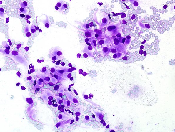
Washington University Experience | NEOPLASMS (MENINGIOMA) | Rhabdoid | 5F Meningioma, atypical w rhabdoid (Case 5) Wright 1.jpg
A Wright stain shows prominent perinuclear staining of the rhabdoid cells. ---- Ancillary data (not shown). Glial fibrillary acidic protein is negative in the tumor cells. Additional markers that are also negative include MART1, pan-cytokeratin and CAM 5.2, CD20, CD45 and HMB-45. S100 shows faint patchy reactivity. ---- FISH shows no evidence of deletion of 1p36 or loss of 14q. Furthermore, there is no evidence of monosomy of chromosome 9/loss of CDKN2A. Finally, there is no loss of the chromosomal arm 22q, the site of the NF2 gene. The histologic and immunohistochemical findings, particularly its H&E appearance, reactivity for EMA and PR receptor, indicate that this tumor represents a meningioma. There are extensive atypical features, including loss of architecture, small cell change and hypercellularity to justify the diagnosis of a grade 2 atypical meningioma. There are predominant rhabdoid features, however we believed that the limited mitotic activity did not warrant the designation of a WHO grade 3 anaplastic meningioma.

