Table of Contents
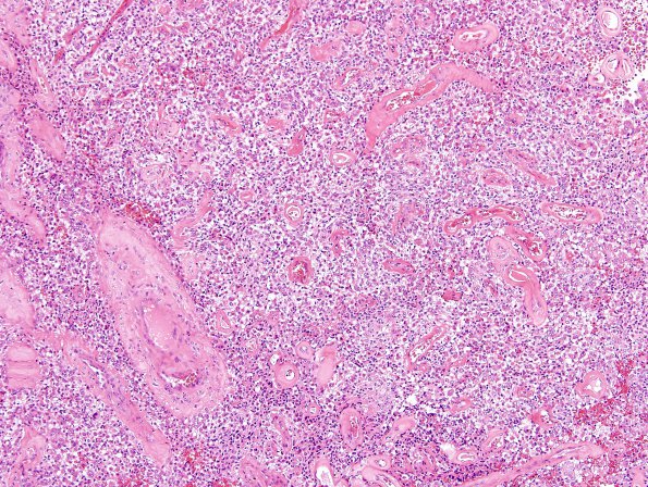
Washington University Experience | NEOPLASMS (MENINGIOMA) | Rhabdoid | 6A1 Meningioma, rhabdoid (2 yo, Case 6) H&E 11 Rhab AP.jpg
Case 6 History ---- The patient was a two year old girl with an intradural, extra-axial right frontal lesion. ---- 6A1-3 Microscopic sections reveal a meningioma with rhabdoid predominant features. Tumor cell density is moderate to high in this neoplasm which does not display necrosis or evidence of brain invasion. Rhabdoid tumor cells contain vesicular nuclei, prominent nucleoli and a round eosinophilic belly of cytoplasm. These cells are arranged in patternless sheets and focally become discohesive. Mitotic activity in these areas is difficult to identify, however, one high powered field does contain three mitotic figures which is worrisome. In addition, there is a focal suggestion of clear cell morphology. "Small cell" morphology (loss of cytoplasm with high N/C ratio) is scattered throughout the tumor tissue as well. Hyalinized blood vessels are a prominent feature in this tumor. Only small foci take on a better differentiated meningothelial appearance; therein, meningothelial whorls are identified.

