Table of Contents
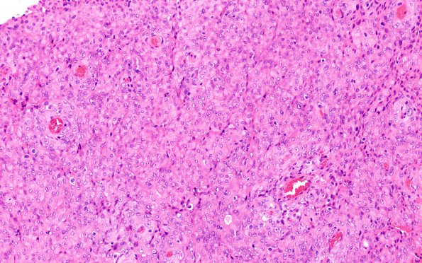
Washington University Experience | NEOPLASMS (MENINGIOMA) | Rhabdoid | 7A1 Meningioma, rhabdoid SP RadioRx (Case 7) H&E 9
Case 7 History ---- The patient was a 44-year-old man with a history of medulloblastoma at the age of two, status post resection followed by chemotherapy and craniospinal radiation. He subsequently developed multiple meningiomas, and is status post approximately nine surgeries and four courses of radiation therapy. These histomorphologic and immunohistochemical findings (particularly the mitotic index in excess of 20) support the diagnosis of anaplastic meningioma with brain invasion, WHO grade 3, likely radiation induced. ---- 7A1-5 Largely arranged in sheets, the tumor cells have atypical round-to-oval nuclei, fairly prominent nucleoli, moderate amounts of cytoplasm, frequent intracytoplasmic whorls, and, often, poorly-defined cell borders. Foci of necrosis are present. (H&E)

