Table of Contents
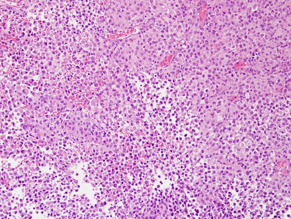
Washington University Experience | NEOPLASMS (MENINGIOMA) | Rhabdoid | 8A1 Meningioma, rhabdoid (Case 8) H&E 21.jpg
Case 8 History ---- The patient was a 54 year old man who presented with a right temporal lesion, originally suspected of representing either a low-grade glioma or a ganglioglioma (1998). A recurrence was resected in 2000. ---- Sections from the original needle biopsy in 1998 were reviewed and revealed a hypercellular neoplasm with spindled and epithelioid morphology. Mitotic figures are identified and no native brain parenchyma is seen. Although the sections suggest a vaguely "glial" appearance, the tissue preservation is suboptimal. Likewise, vaguely "ganglion-like" cells with eccentric nuclei and abundant cytoplasm are best characterized retrospectively as rhabdoid. ---- 8A1-7 Based on the appearance of the subsequent biopsy, the diagnosis of anaplastic rhabdoid meningioma is favored. Neoplastic cells are characterized by abundant eccentric cytoplasm and large nuclei with prominent nucleoli, The mitotic index is quite brisk and there are extensive areas of tumoral necrosis. In a single section, a lower grade appearing component is identified. In this region, the tumor takes on a meningothelial appearance with whorling architecture.

