Table of Contents
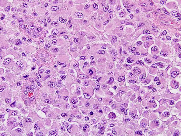
Washington University Experience | NEOPLASMS (MENINGIOMA) | Rhabdoid | 8A7 Meningioma, rhabdoid (Case 8) H&E 17.jpg
Higher magnification additional image of this neoplasm. (H&E) ---- Results of immunostained sections demonstrate strong diffuse immunoreactivity for vimentin which highlights paranuclear inclusions. Patchy expression of EMA is also appreciated. There is no tumoral expression for glial fibrillary acidic protein (GFAP), synaptophysin, desmin or smooth muscle actin. The morphologic and immunohistochemical features are consistent with the diagnosis of rhabdoid meningioma with anaplastic features, WHO grade 3. ---- Fluorescence in situ hybridization (FISH) studies were performed from paraffin embedded material from both the 1998 and 2000 biopsies. Both specimens revealed a deletion for the NF2 region on 22q12, whereas the normal two copies (disomy) of BCR (22q11) were appreciated. This suggests a 22q deletion that includes NF2 and excludes the BCR region. Given the proximity of the BCR gene to the INI1 gene implicated in most primary rhabdoid tumors, deletions have been demonstrable in the great majority of atypical teratoid/rhabdoid tumors utilizing this locus. Therefore, the genetic profile is most consistent with a meningioma that has undergone rhabdoid transformation rather than a primary rhabdoid tumor of the CNS.

