Table of Contents
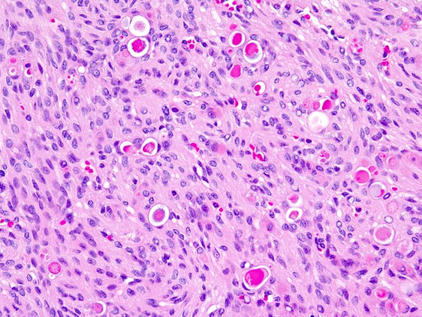
Washington University Experience | NEOPLASMS (MENINGIOMA) | Rhabdoid | 9A1 1Meningioma, secretory (Case 9) 3.jpg
Case 9 History ---- The patient was a 65 year old woman with a right frontal meningioma measuring 5 x 4 cm with a large area of surrounding edema. Operative procedure: Resection. ---- 9A1,2 This neoplasm is composed of large aggregates and fascicles of fibrous and meningothelial cells. Many of the meningothelial cells have eosinophilic, abundant cytoplasm and eccentric nuclei, a rhabdoid morphology. Mitotic figures, up to 2/10HPF, are present. In addition, there are foci of small cell change, sheeting, and macronuclei. Numerous eosinophilic, intralumenal globules are present, highlighted by a PAS with diastase stain as expected in the secretory variant of meningioma. Immunohistochemical stain for cytokeratin and monoclonal carcinoembryonic antigen show positive reactivity in the cells forming these globules.

