Table of Contents
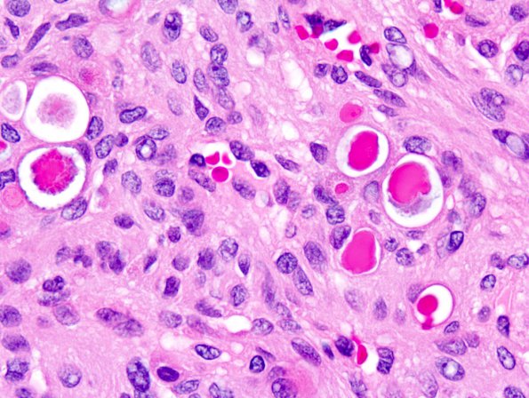
Washington University Experience | NEOPLASMS (MENINGIOMA) | Secretory | 15B4 Meningioma, secretory (Case 15) 4.jpg
Higher magnification tumor image (H&E) ---- Ancillary studies (not shown): Immunohistochemically, the tumor showed positive reactivity to vimentin and EMA and was negative for cytokeratin. The stain for Ki-67 showed a low proliferation index. Several features in this neoplasm, including focal rhabdoid morphology, sheeting, small cell change, and macronuclei suggests the potential for more aggressive clinical behavior. Intraluminal globules are present, highlighted by a PAS - diastase stain. Immunohistochemical stain for cytokeratin and monoclonal CEA show positive reactivity in the cells forming the globules. The morphological and immunohistochemical features are those of a secretory meningioma.

