Table of Contents
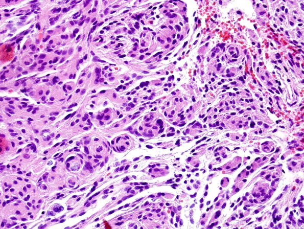
Washington University Experience | NEOPLASMS (MENINGIOMA) | Transitional | 10A Meningioma (Case 10) H&E 1
Case 10 History ---- The patient is a 60 year old woman with progressive visual loss in left eye and a tuberculum sellar base mass on MRI. Operative findings of a soft, purple-red dural base mass. ---- 10A There is mild nuclear pleomorphism, with the majority of tumor cells containing round to oval nuclei and abundant eosinophilic cytoplasm. Many of the nuclei have clear holes or nuclear pseudo inclusions. Atypical features are not noted. Mitotic figures are not easily identified and there is no tumor necrosis.

