Table of Contents
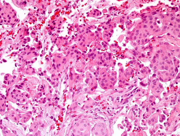
Washington University Experience | NEOPLASMS (MENINGIOMA) | Transitional | 12A Meningioma (Case 12) H&E 1.jpg
Case 12 History ---- The patient is a 40-year-old woman with unilateral vision changes. MRI showed a small enhancing lesion near the right optic foramen and olfactory groove suggestive of a small meningioma affecting the right optic nerve. Operative procedure: Right craniotomy for tumor excision. ---- 12A Sections of the resected lesion show a meningothelial neoplasm in which tumor cells are arranged in cohesive groups separated by fibrous intervening tissue. Tumor nodules have round to oval nuclei, occasional intranuclear pseudoinclusions and longitudinal nuclear grooves. There is no appreciable mitotic activity and no subjective features of atypia.

