Table of Contents
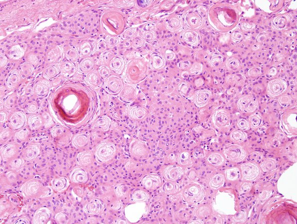
Washington University Experience | NEOPLASMS (MENINGIOMA) | Transitional | 14A Meningioma (Case 14) H&E 9
Case 14 History ---- The patient is a 52 year old woman with several episodes of neurological symptoms. MRI of the brain shows homogenously enhancing extra axial masses in the right frontal region, along the greater wing of the right sphenoid bone, and based on the planum sphenoidale. Operative procedure: Right frontal-pterional craniotomy and removal of right frontal convexity meningioma, right sphenoid wing meningioma, and tuberculum sella meningioma with use of intraoperative MRI. ---- 14A The various submitted specimens all show the presence of meningothelial neoplasms. The specimens show a neoplasm consisting of cells that form prominent nodules, whorls, and psammoma bodies. The neoplastic cells have round to ovoid nuclei, mild nuclear membrane irregularities, variable chromatin ranging from open to stippled, and eosinophilic cytoplasm. Intra-nuclear cytoplasmic 'pseudo'-inclusions are present. There are scattered areas of cells with increased nuclear to cytoplasmic ratios indicative of small cell change. Mitoses are not identified. Other atypical features are not observed (i.e. sheeting, patternless growth, spontaneous necrosis, prominent macronucleoli, and increased mitotic activity).

