Table of Contents
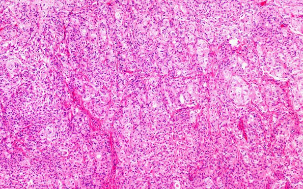
Washington University Experience | NEOPLASMS (MENINGIOMA) | Transitional | 1A1 Meningioma, transitional (Case 1) A7 H&E 10X
Case 1 History ---- The patient is a 55-year-old woman presenting with right vision loss with imaging showing a right intracranial fossa
meningioma. Operative procedure: Right craniotomy tumor excision; right transcranial orbitotomy; right implant placement outside muscle cone. ----
1A1-3 Hematoxylin and eosin stained sections show a meningothelial neoplasm with overwhelmingly whorled architecture. The tumor cells are arranged in lobules with smaller interspersed whorls and psammoma bodies. The tumor cells have abundant eosinophilic cytoplasm and round eccentric nuclei with vesicular chromatin. A subpopulation of cells have rhabdoid-like morphology. Other areas have a fibrous appearance. Mitoses are overall rare, but hot spots reach up to 3/10HPF. There are large areas of spontaneous necrosis. Additional atypical histologic features are not identified. There are multiple minute foci of brain invasion along the nodular surface of the tumor (A4).

