Table of Contents
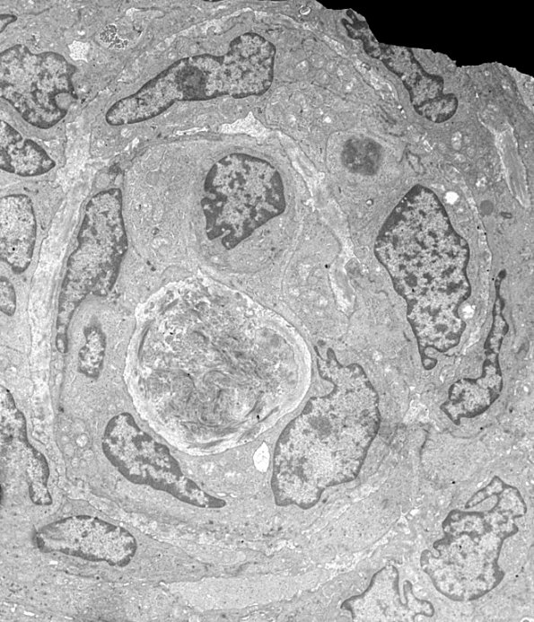
Washington University Experience | NEOPLASMS (MENINGIOMA) | Transitional | 3C Meningioma (Case 3) EM 2 - Copy (2)
Ultrastructure of a whorl. (electron micrograph) ---- Not shown: EMA staining shows positive membranous staining within the tumor cells. Progesterone receptor shows patchy positive nuclear staining within the tumor. Proliferation index (Ki-67) is focally increased up to 7.6%. Based on histomorphological features, and immunostaining profile, this neoplasm is most consistent with a meningioma with focal atypical features, WHO grade 2.

