Table of Contents
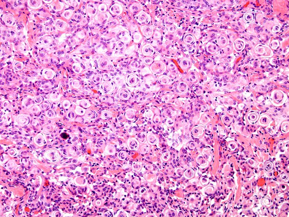
Washington University Experience | NEOPLASMS (MENINGIOMA) | Transitional | 7A1 Meningioma (WHO I, Case 7) H&E 1
Case 7 History ---- The patient is a 36 year old woman with new onset of seizures. MRI showed a dural based uniformly enhancing extra-axial mass arising in the left sphenoid wing and anterior clinoid process. Operative procedure: Craniotomy with tumor excision. ---- 7A1-3 Sections of the biopsy and excision material show a cellular tumor characterized by a whorled architecture as well as numerous tightly formed nodules of spindled cells. The cells have minimal nuclear atypia with stippled chromatin and inconspicuous nucleoli. Scattered psammoma bodies are identified. Mitotic figures are rare. There is no evidence of hypercellularity, small cell change, sheeted architecture, or brain invasion.

