Table of Contents
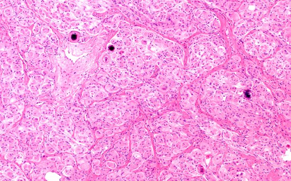
Washington University Experience | NEOPLASMS (MENINGIOMA) | Transitional | 8A1 Meningioma, WHO I whorls (Case 8) H&E 2
Case 8 History ---- The patient is a 51 year old female with an enhancing extra-axial mass along the inner table of the left aspect of the frontal bone that exerts signet and mass effect on the left cerebral hemisphere. There is a dural tail and hyperostosis of the frontal bone in this region. Operative procedure: Excision. ---- 8A1,2 Hematoxylin and eosin stained sections of the "brain tumor" show several fragments of a dural based spindle cell neoplasm. The tumor cells have ovoid nuclei, inconspicuous nucleoli, occasional intranuclear clearing and pseudoinclusions, and moderate amounts of eosinophilic cytoplasm. Mitotic figures are rare, estimated at less than 1/10HPF. Other atypical features, e.g. sheeting architecture, small cell change, macronucleoli, spontaneous necrosis, and hypercellularity, are not appreciated.

