Table of Contents
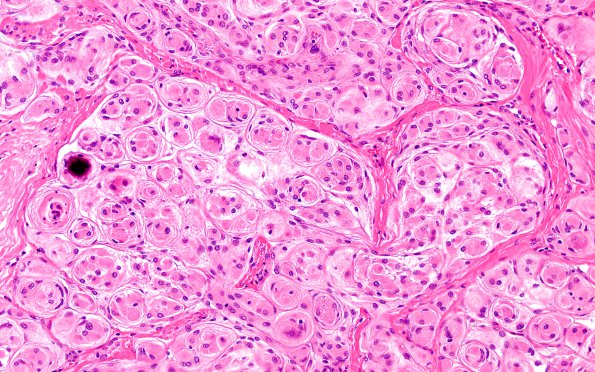
Washington University Experience | NEOPLASMS (MENINGIOMA) | Transitional | 8A2 Meningioma, WHO I whorls (Case 8) H&E 1
Hematoxylin and eosin stained sections of the "brain tumor" show several fragments of a dural based spindle cell neoplasm. The tumor cells have ovoid nuclei, inconspicuous nucleoli, occasional intranuclear clearing and pseudoinclusions, and moderate amounts of eosinophilic cytoplasm. ---- Not shown: The tumor cells are positive for PR. Ki-67 highlights a proliferation index focally up to 3.9% in the tumor cells. The histopathological and immunohistochemical findings are consistent with meningioma, WHO grade I.

