Table of Contents
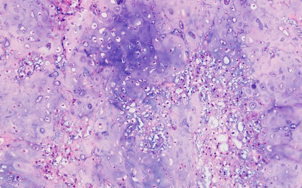
Washington University Experience | NEOPLASMS (MESENCHYMAL, NON-MENINGOTHELIAL) | Chordoma | 13A1 Chondroid chordoma (Case 13) H&E 4
Case 13 History ---- The patient was a 73-year-old man who presented with diplopia. Brain MRI showed a 3.6 cm heterogeneously enhancing midline clivus lesion with bubbly T2 appearance. Operative procedure: Endoscopic endonasal transsphenoidal approach to the middle and posterior fossa skull base, Stealth frameless stereotaxic resection of middle and posterior fossa extradural skull base tumor. ---- 13A1-3 Routine H&E stained sections of both specimens show a neoplasm composed of large epithelioid cells arranged in cords and nests. The tumor cells have round nuclei with abundant eosinophilic cytoplasm, some of which contain multiple clear physaliphorous cells. Areas with pseudo-hyaline cartilaginous matrix are also seen.

