Table of Contents
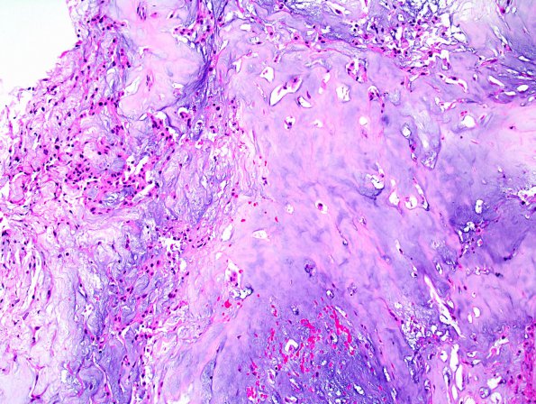
Washington University Experience | NEOPLASMS (MESENCHYMAL, NON-MENINGOTHELIAL) | Chordoma | 15A Chordoma, chondroid (Case 15) 2.jpg
Case 15 History ---- The patient was a 68 year old man with a 3 x 2.8 cm contrast enhancing mass arising from the sella and clivus. A biopsy of the pituitary region was performed. ---- 15A Sections reveal multiple fragments of anterior pituitary parenchyma, as well as fragments of bone involved by a low to moderately cellular neoplasm with mixed epithelioid and chondroid features. In some areas, the tumor is arranged in ribbons of epithelioid cells with moderate quantities of eosinophilic cytoplasm and occasional clear bubbly vacuoles (physaliphorous cells). Other areas blend into regions of hyaline cartilage. The tumor has a permeative growth pattern in between bony spicules. Mitotic figures are hard to find and there is no evidence of tumor necrosis. (H&E)

