Table of Contents
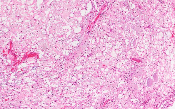
Washington University Experience | NEOPLASMS (MESENCHYMAL, NON-MENINGOTHELIAL) | Chordoma | 17A Chordoma (Case 17) H&E 10X - Copy (3)
Case 17 History ---- The patient was a 32-year-old man who presented with headaches and vision problems. MRI showed an anterior skull base mass, with minimal enhancement. Imaging differentials included epidermoid cyst, chordoma and chondrosarcoma. Operative procedure: Craniotomy with resection of mass. ---- 17A Hematoxylin and eosin stained sections show a low-grade neoplasm. Myxoid matrix separated with fibrous septa showing epithelioid cells arranged in cords, clusters, and small sheets. There are abundant multivacuolated “physaliphorous” cells with clear foamy cytoplasm.

