Table of Contents
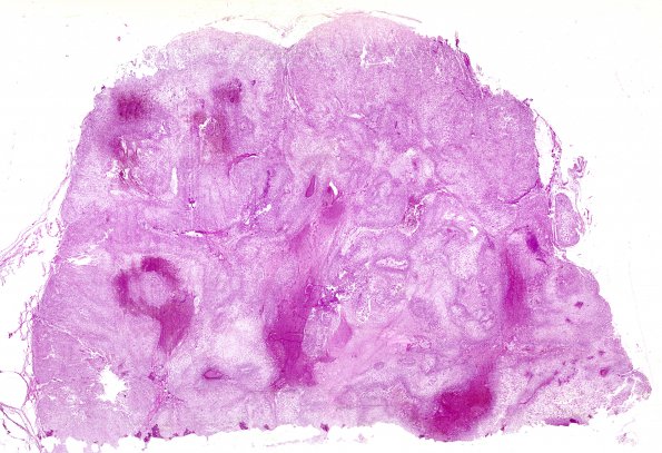
Washington University Experience | NEOPLASMS (MESENCHYMAL, NON-MENINGOTHELIAL) | Chordoma | 19A1 Chordoma, clivus (AFIP TS M9665 set Slide # 83, RES image) H&E whole mount
Case 19 History (AFIP TS M9665 set Slide 83, RES image) ---- The patient was a 53-year-old woman who was admitted to the hospital with a history of intermittent headaches over the previous 4- to 5-year period. The headaches had increased in frequency and duration in the 4 months prior to admission and had localized behind the right eye. At that time the patient had some visual difficulty. Examination revealed constriction of the left temporal field, diplopia, and weakness of conversion of the left eye. The results of the neurologic examination were otherwise unremarkable. X-ray examination showed enlargement and erosion of the sella turcica, suggesting a pituitary tumor. X-ray therapy was elected and a course given. Her visual difficulties, however, continued to increase in severity, and she was readmitted to the hospital, and a tumor of the clivus was removed. The patient received several additional courses of radiation therapy, but her condition grew worse, and she expired 26 months after her first admission to the hospital. 19A1 2 The tumor is strongly nodular and composed of typical chordoma elements (H&E).

