Table of Contents
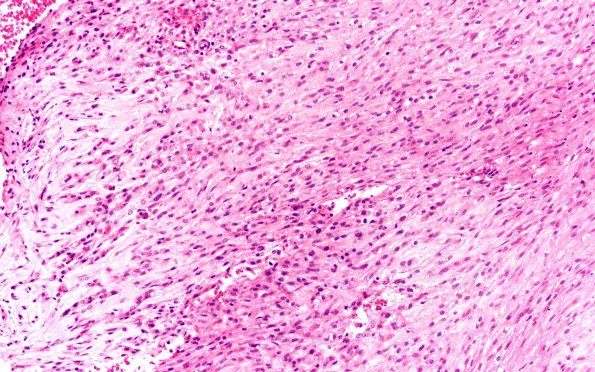
Washington University Experience | NEOPLASMS (MESENCHYMAL, NON-MENINGOTHELIAL) | Chordoma | 21A3 Chordoma (Case 21) 4X spindle cell area H&E 20X
This area is taken from the arrowhead region of image 21A1. There are small and large foci with high cellularity and no intercellular substance, where the neoplastic cells are spindling and arranged in a storiform pattern. Although they show nuclear atypia in these areas, they are not highly pleomorphic and they do not show significant mitotic activity. The morphology of these areas is consistent with early dedifferentiation of the tumor to a spindle cell sarcoma but it is not compellingly malignant. Focal areas of coagulative necrosis are also seen.

