Table of Contents
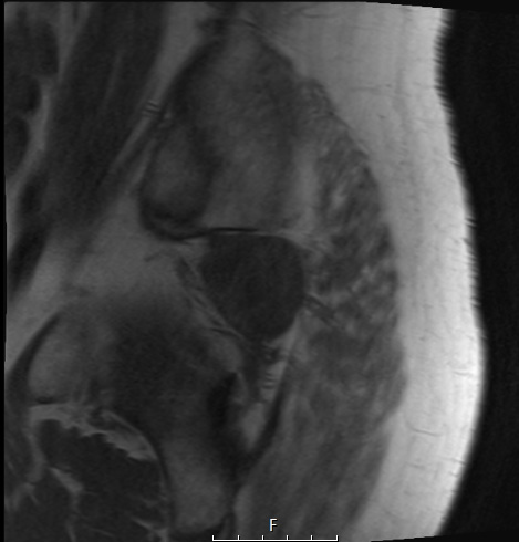
Washington University Experience | NEOPLASMS (MESENCHYMAL, NON-MENINGOTHELIAL) | Chordoma | 2A1 Chordoma (Case 2) T1 no C - Copy
Case 2 History ---- The patient was a 68 year old man who presented with symptoms of radiculopathy and was found to have a large sacral mass. A biopsy performed and evaluated at an outside institution, but not reviewed at Washington University, yielded a diagnosis of chordoma. ---- 2A1-4 MRI of the pelvis showed a large (~11 x 13 cm) well-circumscribed, lobulated mass with mixed T1 and T2 signals and heterogeneous enhancement, occupying the sacrum with extension through the coccyx into soft tissues anterior and posterior to the coccyx and sacrum, and into the sciatic notch regions bilaterally, right more so than left. Clinical/radiological diagnosis: Sacrococcygeal chordoma. Operative procedure: Posterior lumbar decompression L4/5 and sacral tumor resection. ---- 2A1,2 T1 weighted image is hypointense (2A1), increasing in intensity with contrast (2A2)

