Table of Contents
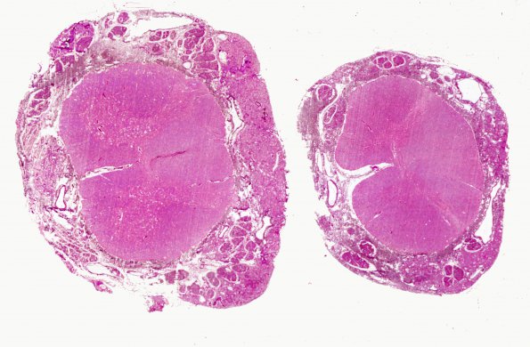
Washington University Experience | NEOPLASMS (MESENCHYMAL, NON-MENINGOTHELIAL) | Hemangioblastoma | 10B3 Hemangioblastoma (Case 10) Retic WM
10B3,4 A whole mount of spinal cord and low magnification image of the ventral roots running through the specimen. Tumor intimately envelopes, but does not invade, both cranial and spinal nerves. Nerve fiber loss is seen focally in the cranial nerves and prominently in the dorsal roots. Secondary dorsal column degeneration is seen.

