Table of Contents
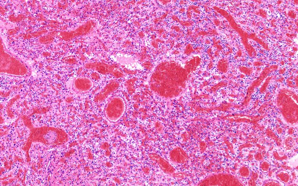
Washington University Experience | NEOPLASMS (MESENCHYMAL, NON-MENINGOTHELIAL) | Hemangioblastoma | 13A1 Hemangioblastoma (Case 13) H&E 2
Case 13 History ---- The patient is a 22-year-old man with history of traumatic brain injury with 6 months of progressive dizziness, discoordination, occipital headache, and diplopia, with brain MRI demonstrating a 5.6 cm cystic cerebellar mass with a 1.3 cm mural nodule, concerning for pilocytic astrocytoma versus hemangioblastoma. Operative procedure: Posterior fossa craniotomy. ---- 13A1,2 Sections of the cerebellar lesion show a neoplasm consisting of numerous small vascular spaces surrounded by bland, epithelioid stromal cells with eosinophilic-to-vacuolated cytoplasm. Mitotic figures and necrosis are not identified. Compare 13A1 and 13A2. One area appears fairly traditional and the other area is composed solely of stromal cells and might be considered to mimic clear cell carcinoma. The tumor shows a well-defined border with the surrounding cerebellar cortex, which shows loss of Purkinje neurons, likely reactive in nature.

