Table of Contents
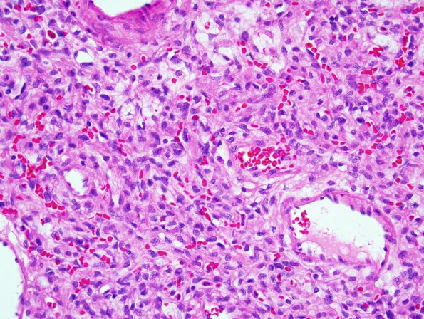
Washington University Experience | NEOPLASMS (MESENCHYMAL, NON-MENINGOTHELIAL) | Hemangioblastoma | 18A Hemangioblastoma (Case 18) 1.jpg
Case 18 History ---- The patient is a 72 year old woman who had a prior history of "cerebellar glioma" in 1985. Twenty-three years later she presented with a cerebellar tumor, consistent with tumor recurrence. ---- 18A Sections reveal a relatively well demarcated markedly vascular tumor. In between the capillaries and larger thin-walled vessels are numerous small tumor cells with clear foamy cytoplasm and moderate nuclear pleomorphism. The adjacent brain parenchyma shows hemosiderin-laden macrophages and gliosis with scattered Rosenthal fibers. Mitotic figures are infrequent and there is no definite evidence of tumor necrosis.

