Table of Contents
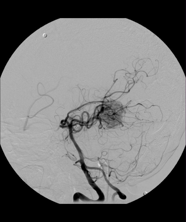
Washington University Experience | NEOPLASMS (MESENCHYMAL, NON-MENINGOTHELIAL) | Hemangioblastoma | 20A1 Hemangioblastoma (Case 20) Angiogram R Vert obl 2 - Copy
Case 20 History ---- The patient is a 32 year old man with a cystic posterior fossa mass with a large enhancing solid portion on MRI, status post embolization. Operative procedure: Craniotomy for vascular mass in superior vermis/pineal region. ---- 20A1 This image shows an angiogram following vertebral artery injection.

