Table of Contents
Washington University Experience | NEOPLASMS (MESENCHYMAL, NON-MENINGOTHELIAL) | Hemangioblastoma - Von Hippel Lindau | 1A1 Hemangioblastoma (Case 1, Von Hippel Lindau) CT 27 copy - Copy
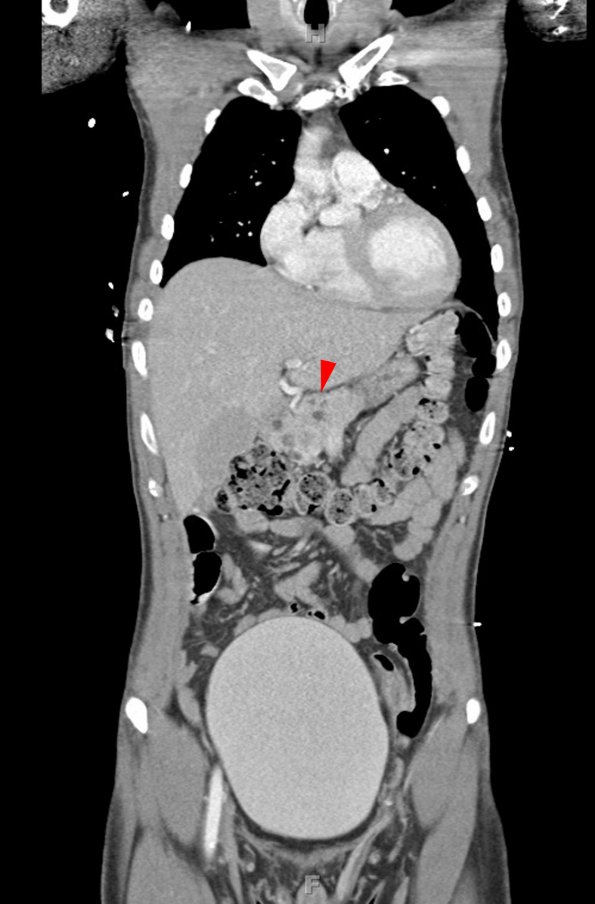
Case 1 History ---- The patient is a 27-year-old man with Von Hippel Lindau syndrome who recently developed right sided headaches and vomiting. Abdominal CT scan showed bilateral cystic renal parenchymal lesions consistent with renal cell carcinoma. Head CT scan showed a 5.5 x 3.8 x 3.7 cm hypodense mass with markedly irregular peripheral enhancement and mural nodularity that expanded the cerebellar vermis and extended inferiorly to the cisterna magna at the level of C2. There was associated proximal anterior cord compression. Operative procedure: Suboccipital craniotomy for tumor resection. ----1A1-5 Whole body CT sections of a patient with the classical appearance of VHL.
1A1,2 These images show a longitudinal (1A1) and cross (1A2) section at the level of the pancreas exhibiting numerous cysts (arrowhead, 1A1).

