Table of Contents
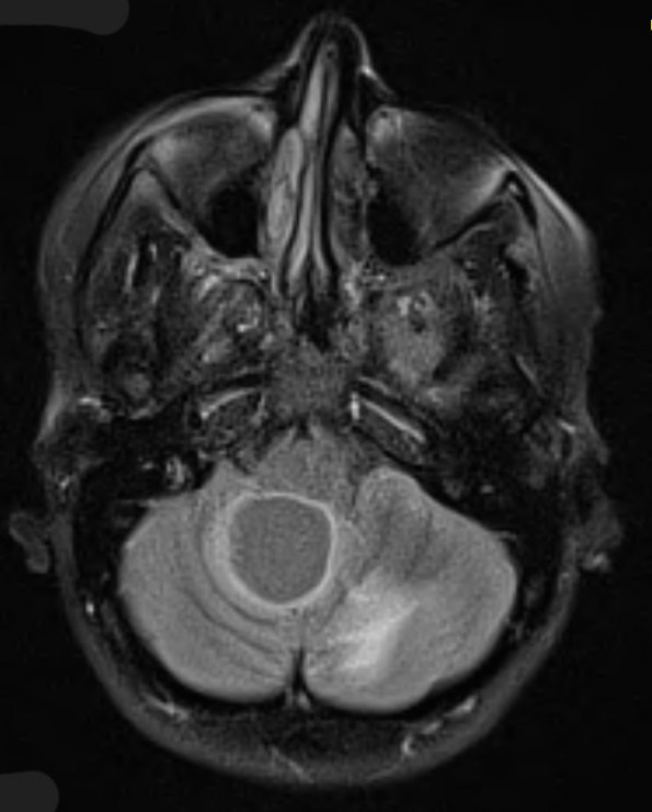
Washington University Experience | NEOPLASMS (MESENCHYMAL, NON-MENINGOTHELIAL) | Hemangioblastoma - Von Hippel Lindau | 3A1 Hemangioblastoma (VHL, Case 3) FLAIR - Copy
Case 3 History ---- The patient is a 27 year old woman who presented with headaches and increased episodes of dizziness. MRI of the brain shows a cystic right cerebellar lesion containing an enhancing mural nodule and an additional enhancing small spherical lesion in the contralateral (left) cerebellar hemisphere. The primary differential consideration is hemangioblastoma. A chest, abdomen, and pelvis CT showed multiple pancreatic cysts and a low attenuation right renal lesion. She has a strong family history of Von Hippel-Lindau disease. Operative procedure: Posterior fossa craniotomy with stealth guidance. ---- 3A1 This FLAIR scan shows a focus of hyperintense cystic rim in the right cerebellar hemisphere and a margin of an additional lesion in the left cerebellar hemispheric.

