Table of Contents
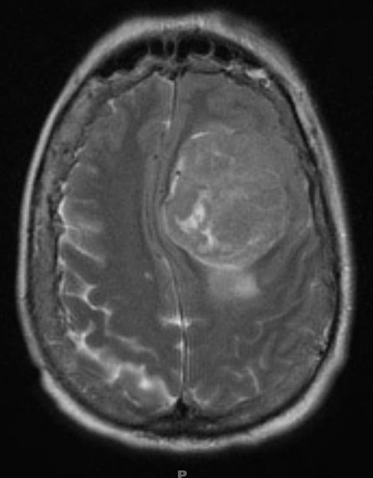
Washington University Experience | NEOPLASMS (MESENCHYMAL, NON-MENINGOTHELIAL) | Solitary Fibrous Tumor (SFT) | 12A Hemangiopericytoma, Grade III (Case 12) T2 - Copy
Case 12 History ---- The patient was a 65 year old woman with slowly progressive cognitive decline and more recent development of aphasia. Radiographic imaging without contrast revealed a 6.6 cm left frontal extra-axial mass, isointense to gray matter on T1 and T2-weighted imaging, without evidence of hemorrhage or calcification. The tumor was not embolized prior to resection. Operative procedure: Craniotomy for resection of tumor.---- This large tumor is well seen on this T2-weighted MRI scan.

