Table of Contents
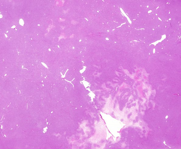
Washington University Experience | NEOPLASMS (MESENCHYMAL, NON-MENINGOTHELIAL) | Solitary Fibrous Tumor (SFT) | 12C1 Hemangiopericytoma, Grade III (Case 12) H&E whole mount
12C1-6 The tumor consists of hypercellular sheets of cells with a high nucleus-to-cytoplasm ratio, modest eosinophilic cytoplasm, oval nuclei with irregular contours and speckled chromatin, and distinct eosinophilic nucleoli. Mitotic figures are frequent >5/10HFT The tumor tissue is quite vascular, with a dense array of minute capillaries, small vessels appearing as polygonal spaces or slits, and larger vessels appearing as branched staghorn-like figures. Interrupting this sheet-like growth pattern are areas of geographic necrosis, ranging over a centimeter in greatest dimension. Histochemically stained sections show reticulin fibers surrounding larger vessels, and investing individual tumor cells with variable but considerable frequency.

