Table of Contents
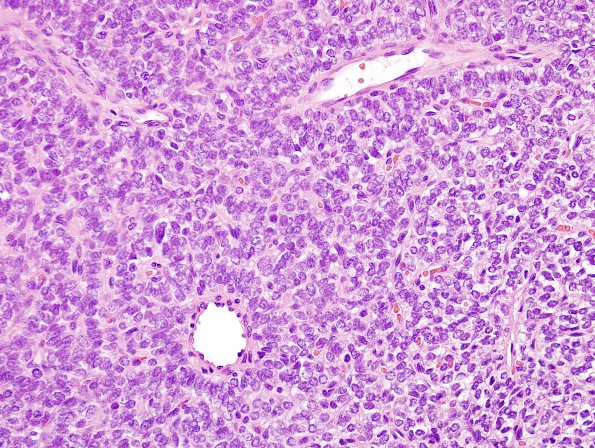
Washington University Experience | NEOPLASMS (MESENCHYMAL, NON-MENINGOTHELIAL) | Solitary Fibrous Tumor (SFT) | 14G1 2nd resection Hemangiopericytoma, metastatic (Case 14) H&E 1.jpg
2014 Surgical specimen ---- Case 14 History ---- The same patient as the previous resection is a now 49-year-old woman with a history of SFT, WHO grade III, in the left middle fossa resected in 2010. The patient's tumor was initially diagnosed in 2007, and she has undergone multiple gamma knife surgeries in 2007, 2010, 2011, and January 2014. The patient now presents with left arm pain. MR imaging performed in September 2014 shows an enhancing extramedullary intradural lesion at the C5-C6 intervertebral disc level, occupying the left lateral aspect of the spinal canal and displacing the spinal cord anteriorly. Of note, the patient has known, multiple lesions intracranially, most of which involve the left cerebral hemisphere. A whole body PET scan was performed on September 2014, which showed a lytic lesion with associated cortical destruction within the intertrochanteric region of the right proximal femur, likely representing osseous metastasis. Operative procedure: C4-C6 laminectomy for excisional biopsy of the intradural/extramedullary lesion. ---- 14G1,2 Routine hematoxylin-and-eosin stained sections of the "extramedullary intradural tumor" show a well-circumscribed, highly cellular tumor with rich vascularity. Some of the vessels have a characteristic "staghorn" appearance. The neoplastic cells have scant amounts of eosinophilic cytoplasm, and contain round to oval nuclei with coarse chromatin and rare, small nucleoli. The nuclei are moderately pleomorphic. Mitotic figures are rare (less than 1 per 10 high powered fields). Necrosis is not identified.

