Table of Contents
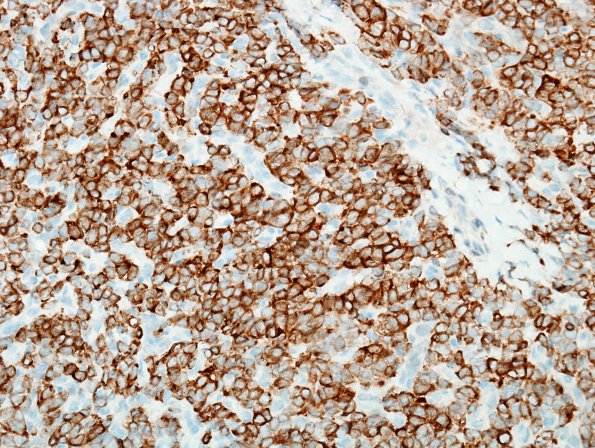
Washington University Experience | NEOPLASMS (MESENCHYMAL, NON-MENINGOTHELIAL) | Solitary Fibrous Tumor (SFT) | 14H2 2nd resection Hemangiopericytoma, metastatic (Case 14) Bcl-2 1.jpg
A CD34 stain highlights both the rich vasculature and scattered individual tumor cells. ---- A bcl-2 immunostain highlights a majority of cells with cytoplasmic distribution. ---- Ancillary findings (not shown): A reticulin stain highlights the vasculature and demonstrates focally increased reticulin framework around individual cells. A Ki-67 immunostain demonstrates an index of proliferation of approximately 2.5%.The 2014 specimen was compared with the patient's previous 2010 resection. Although the morphologic features are similar, the previously resected material shows a higher mitotic index as well as a higher index of proliferation. However, given the presence of multiple intracranial lesions and a new bony metastasis to the femur, this tumor is best classified as a metastatic SFT.

