Table of Contents
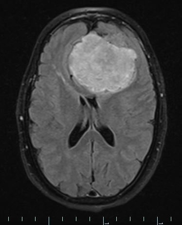
Washington University Experience | NEOPLASMS (MESENCHYMAL, NON-MENINGOTHELIAL) | Solitary Fibrous Tumor (SFT) | 1A1 HAP-SFT, WHO 3 (Case 1) FLAIR 1 - Copy
Case 1 History ---- The patient was a 59-year-old woman with a 2-3 week history of altered mental status and lethargy. ED workup pointed to renal failure and pancreatitis and she underwent dialysis, during which time she experienced a seizure. Subsequent brain MRI showed an extra-axial, lobular, complex, heterogeneously enhancing mass measuring 6.6 x 6 x 5.8 cm which compressed the anterior left frontal lobe, and extends beneath the anterior falx and crossing the midline by 2.3 cm. Operative procedure: Stereotactic guided left frontal temporal craniotomy for tumor resection. ---- 1A1,2 MRI showed an enormous mass with FLAIR and T1-weighted contrast enhanced scans.

