Table of Contents
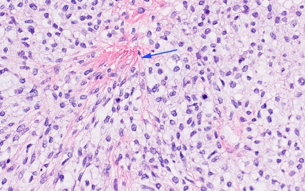
Washington University Experience | NEOPLASMS (MESENCHYMAL, NON-MENINGOTHELIAL) | Solitary Fibrous Tumor (SFT) | 1B3 HAP-SFT, WHO 3 (Case 1) H&E 4 copy
H&E showed a high-grade mesenchymal neoplasm with a heterogeneous appearance. The predominant portion of the tumor consists of relatively hypocellular, lobulated myxoid areas, with tumor cells featuring pleomorphic ovoid nuclei with occasional nuclear scalloping, coarse to vesicular chromatin, inconspicuous nucleoli, and indistinct cell borders. Throughout the myxoid background there are plaque-like collagen deposits with a brightly eosinophilic appearance, so called “amianthoid fibers” (arrow 1B3). Ectatic, thin-walled, branching vessels (“staghorn vessels”) are also seen throughout the tumor. The second component of the tumor, which is seen in some well-demarcated areas and in others to be intermixed with the myxoid complement, has a hypercellular and more fibrous appearance. Mitotic activity is elevated (8 mitoses/10HPF). There is no evidence of necrosis.

