Table of Contents
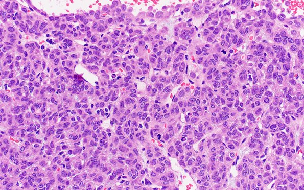
Washington University Experience | NEOPLASMS (MESENCHYMAL, NON-MENINGOTHELIAL) | Solitary Fibrous Tumor (SFT) | 2A3 Hemangiopericytoma, anaplastic (WHO III, Case 2) H&E 2
Routine H&E stains show a dural-based hypercellular neoplasm. The tumor cells have hyperchromatic nuclei, irregular nuclear contours, frequent prominent nucleoli, and moderate amounts of eosinophilic to focally clear cytoplasm. There are many dilated, thin-walled vessels, including some with a staghorn pattern. Mitotic activity reaches 21 mitoses/10HPF. Necrosis is not appreciated.

