Table of Contents
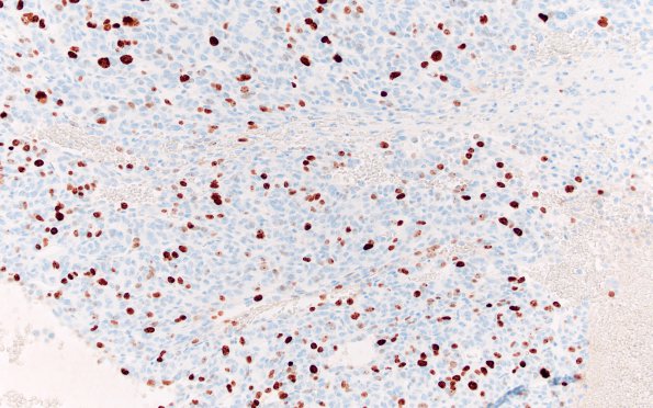
Washington University Experience | NEOPLASMS (MESENCHYMAL, NON-MENINGOTHELIAL) | Solitary Fibrous Tumor (SFT) | 2D Hemangiopericytoma, anaplastic (WHO III, Case 2) Ki67 20X
2D Ki-67 (MIB-1 antibody) highlights a proliferation index of 28.1% in some areas (Ki67 IHC) ---- Ancillary findings (not shown): Tumor cells are focally positive for BCL2, CD56, EMA (punctate, weak), PR, and CD34 (weak). Rare cells show reactivity for synaptophysin. Pankeratin, S100, and GFAP highlight uninvolved brain parenchyma and are non-reactive in the tumor. Smooth muscle actin, ERG, CD34, and reticulin highlight the rich vasculature while EMA highlights occasional arachnoid cap cells. Of note, the tumor has a poor reticulin meshwork lacking pericellular reticular fibers outside of the vascular basement membranes. PHH3 highlights scattered tumor nuclei. Tumor cells are positive for vimentin but negative for HMB-45, CAM 5.2, and chromogranin.
Fluorescence in situ hybridization studies are negative for rearrangement of SS18 or EWSR1 genes and loss of chromosome 22q.
Comment:
The tumor has extensive mitotic activity but fails to show necrosis and diagnosed as an SFT WHO Grade 2.

