Table of Contents
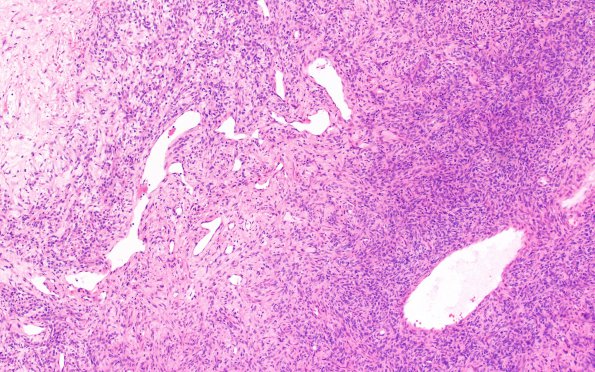
Washington University Experience | NEOPLASMS (MESENCHYMAL, NON-MENINGOTHELIAL) | Solitary Fibrous Tumor (SFT) | 7B1 SFT (Case 7) H&E 20X 1
7B1-3 H&E stained sections show a hypercellular, short spindled cell neoplasm with abundant vasculature that regionally harbors a staghorn appearance. Mitoses are focally increased reaching ~12-14/10 HPF. Patchy hypocellular areas with loose collagen deposition are also present. Scattered apoptotic debris is seen, although definite areas of necrosis are not evident. The tumor shows regional infiltration of the overlying dura through its entire thickness. Intravascular embolization material is present in multiple areas.

