Table of Contents
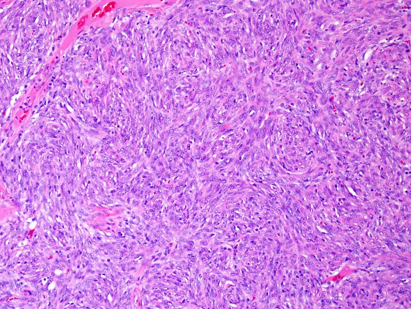
Washington University Experience | NEOPLASMS (MESENCHYMAL, NON-MENINGOTHELIAL) | Solitary Fibrous Tumor (SFT) | 9A1 Hemangiopericytoma (HAP, Original Cervicomedullary site, Case 9) H&E 1.jpg
Case 9 History ---- The patient was a 51 year old man who developed headaches in 2014 that led to the discovery of an enhancing lesion in the region of the medulla. He underwent surgery followed by radiation. In 2015, he developed weakness in his legs and an MRI showed an enhancing mass at T8-T9. Since then, he developed progressive right leg weakness with headaches. Spine imaging from June of 2016 showed cervical and thoracic lesions suggestive of subarachnoid tumor spread. Operative procedure: Resection. ---- 9A1-3 Medullary lesion (2014) Sections of the resected tumor from the cervicomedullary region show a mesenchymal neoplasm with a thin tumor capsule and multiple gaping thin-walled vessels. Tumor cells are arranged predominantly in moderately hypercellular sheets with intersecting, variably oriented fascicles to impart a storiform appearance. Tumor cells show spindled morphology with cytoplasmic vacuolation and nuclei with vesicular chromatin. Mitotic figures are present at 5/10HPF.

