Table of Contents
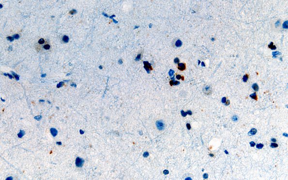
Washington University Experience | NEURODEGENERATION | ALS | 11D2 (Case 11) N12 Motor 60X
An immunohistochemical stain for phosphorylated TDP43 was performed on the motor cortex section and shows cortical neurons with cytoplasmic expression of branched and round inclusions. The motor cortex shows a few granular deposits, some likely within glia. Occasionally lipopigment may complicate the interpretation. (TDP43 IHC) ---- Not shown: Multiple sections of the cerebral cortices (left superior frontal lobe, left superior temporal lobe, left cingulate gyrus, left occipital lobe, and left motor cortex) show a relatively well-maintained complement of neurons without concomitant astrocytic and microglial proliferation. ---- Neuro Final Diagnosis: Amyotrophic lateral sclerosis (ALS) ---- Neuro Diagnosis Comment: TDP43-positive cytoplasmic aggregates are present within motor neurons of the anterior horn, a finding which is documented in the vast majority of patients with ALS. .Additionally, TDP43 proteinopathy is evident within some brainstem motor nuclei despite the neuronal populations there appearing to be intact. Although there are rare cortical neurons within the left motor cortex with TDP43 cytoplasmic expression, the relative paucity of these findings and lack of cortical atrophy do not suggest a significant component of frontotemporal lobar degeneration (FTLD).

