Table of Contents
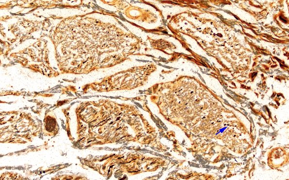
Washington University Experience | NEURODEGENERATION | ALS | 12D3 ALS (Case 12) LS N12 Biels VR 20X copy
Axon loss is minimal in the dorsal roots (12D2) but is nearly complete in the ventral root (12D3). (Bielschowsky) ---- Not shown: Hematoxylin and eosin stained sections of the frontal, occipital, precentral gyrus (N14 and N15), and cingulate gyrus (N16) show no clear evidence of neuronal loss or white matter abnormalities. The motor nuclei of the medulla (N8) appear relatively unremarkable on H&E. Immunohistochemistry (IHC) for phosphorylated transactive response DNA binding protein 43 kDa (TDP43) shows considerable non-specific staining of lipofuscin granules within neurons; nevertheless, one fine skein-like inclusion and multiple thicker inclusions (some globoid, some irregular) are noted in the reticular formation. ---- Neuro Final Diagnosis: Amyotrophic Lateral Sclerosis, Spinal Motor Neuron predominant. ---- Neuro Diagnosis Comment: In addition to these findings of ALS, rare mature amyloid plaques are noted, without clear evidence of neurofibrillary tangle pathology. Although such plaques support the diagnosis ‘Alzheimer disease neuropathologic change,’ the burden of AD pathology is minimal and would not be expected to have any clinical significance.

