Table of Contents
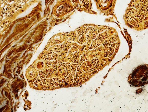
Washington University Experience | NEURODEGENERATION | ALS | 15H2 FTLD-ALS (Case 15) Biels N10 3C (VR) 40X
Ventral roots show diminished numbers of axons compared to dorsal roots. ---- Final Diagnosis- Frontotemporal Lobar Degeneration - Amyotrophic Lateral Sclerosis ---- Neuro Diagnosis Comment: Most individuals do not have a family history of the disease, but in the ~5% that do, several genetic associations are noted. These include mutations in superoxide dismutase (SOD1) which account for 20% of familial cases as well as FUS and TDP43. Some ALS patients have cognitive defects with prominent frontal lobe features very much like those seen in frontotemporal lobar degeneration (FTLD). FTLD is the second most frequent cause of presenile dementia and is a heterogeneous syndrome. While some forms are tau-associated, others have tau-negative, ubiquitin positive inclusions that were found in most cases to be TDP43 positive. This pathological protein was also found to account for the majority of SOD1-negative ALS cases. Most of the remaining ALS and FTLD cases are FUS positive. ALS & FTLD are now considered to represent a pathological spectrum of disease rather than separate processes. In the case of this patient, a 5 year history of clinically diagnosed ALS is supported by the finding of motor neuron loss in the spinal cord, loss of corticospinal tract axons along with evidence of molecular alterations associated with the disease. In both the spinal cord as well as the motor cortex skein-like and occasional round inclusions of TDP43 are noted in neurons within sections stained by TDP43 immunohistochemistry. Occasional cytoplasmic staining in glial cells was also seen. In addition to the classical features of ALS pathology there is atrophy of the frontal lobe. Both neurons and glia showed TDP43 positive inclusions indicating that the pathology represents FTLD. We did not find evidence of neurofibrillary tangles, senile plaques, Pick bodies and cells or cortical Lewy bodies which would indicate additional dementia-related pathologies.(Thanks Grant Kolar MD, PhD for the discussion writeup)

