Table of Contents
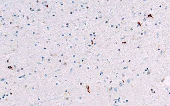
Washington University Experience | NEURODEGENERATION | ALS | 17F2 ALS (Case 17) N16 TDP-43 40X A
There is reactive astrocytosis in the precentral gyrus, modest loss of pyramidal neurons and microvacuolation of the superficial cortex. TDP43 positive cytoplasmic inclusions are also present in the neurons in the precentral gyrus in significant numbers and in scattered glia. (TDP IHC) ---- Not shown: A few Bunina bodies and atrophic neurons were identified in the medullary hypoglossal nuclei. Considering that cortical TDP43 pathology may also be associated with frontotemporal lobar degeneration (FTLD), we also examined the frontal and temporal lobe sections. Although a few TDP43 immunoreactive inclusions, mostly glia, were present in the frontal lobe section, they were fewer than those in the precentral gyrus) and there were none in the temporal lobe/hippocampal formation. In the absence of frontotemporal cortical atrophy, infrequent TDP43 inclusions in frontal and temporal cortex outside of the precentral gyri and clinical lack of dementia, we did not make a diagnosis of FTLD. These findings are consistent with patient's history of amyotrophic lateral sclerosis. ---- Neuro Final Diagnosis: Amyotrophic lateral sclerosis (Motor Neuron Disease-TDP43+ subtype, MND-TDP43)

