Table of Contents
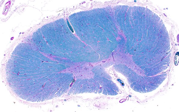
Washington University Experience | NEURODEGENERATION | ALS | 19D (Case 19) 2X LFB-PAS
There is absence of pallor in the lateral and anterior CST (LFB-PAS) ---- Not shown: H&E stained sections of the frontal and precentral gyrus, occipital, and cingulate gyrus show a relatively-well maintained population of cortical neurons. Immunohistochemistry for transactive response DNA binding protein 43 kDa (TDP43) is negative. ---- Neuro Final Diagnosis: Amyotrophic lateral sclerosis ---- Neuro Diagnosis Comment: Post-mortem examination of the central nervous system shows evidence to support the diagnosis of amyotrophic lateral sclerosis (ALS). The brainstem hypoglossal nucleus and motor neurons of the spinal cord demonstrate neuronal loss and atrophic neurons, some of which contain TDP43 immunopositive Bunina bodies and skein-like inclusions. There is minimal, if any, involvement of spinal cord corticospinal tracts. These pathological findings demonstrate a predominance of lower motor neuron pathology and are consistent with the given clinical history ALS

