Table of Contents
Washington University Experience | NEURODEGENERATION | ALS | 1D2 ALS (Case 1) N12 Biels 4X copy
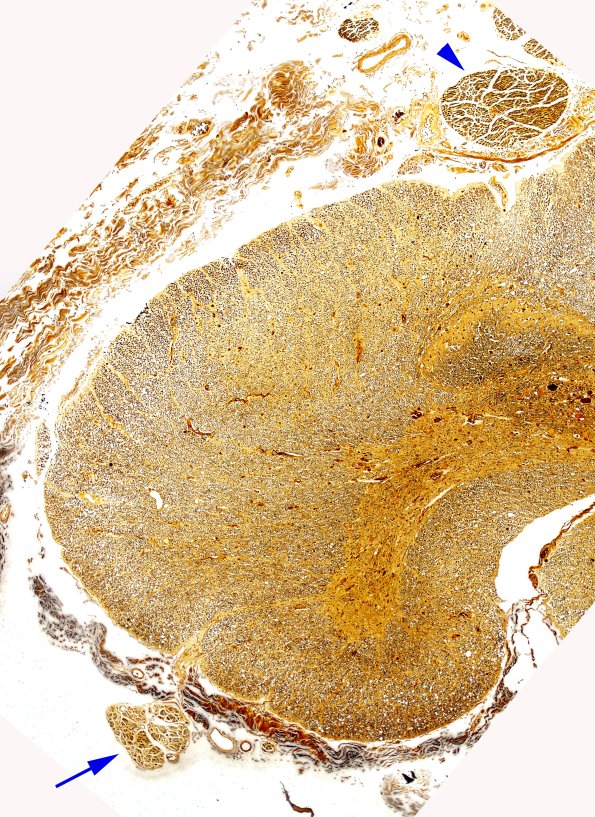
This low magnification image of the thoracic cord specimen stained for myelin shows lateral corticospinal pallor and tissue loss. The parallel loss of myelin and axons is axon degeneration, not demyelination. Notice the dorsal columns are well maintained. Notice the relative size difference between dorsal roots and depopulated ventral roots.

