Table of Contents
Washington University Experience | NEURODEGENERATION | ALS | 7H3 ALS (Case 7) N12 DC 20X
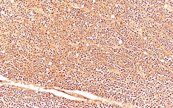
Higher magnification views of axons composing the dorsal columns. (Bielschowsky) ---- Not shown: Sections of the medulla show mild neuronal dropout and reactive astrocytosis in the hypoglossal nucleus. A TDP43 immunostain demonstrates scattered neurons with cytoplasmic staining (inclusions and pre-inclusions), most notably in the regions corresponding to the hypoglossal nucleus and nucleus ambiguus. ---- Neuro Final Diagnosis: Amyotrophic lateral sclerosis with frontotemporal lobar degeneration (ALD-FTLD), characterized by TDP43 proteinopathy involving the spinal cord, brain stem, and cerebral cortices. ---- Neuro Diagnosis Comment: The decedent was noted clinically to have bulbar predominant ALS, which is consistent with evidence of neuronal dropout in the hypoglossal nucleus, as well as the TDP43 inclusions that are evident in the oculomotor nucleus and nucleus ambiguus. Furthermore, TDP43 inclusions are present within many of the remaining anterior horn neurons, as well as many neurons within the cerebral cortices. The latter finding is characteristic of frontotemporal lobar degeneration (FTLD), a pathology often seen in conjunction in ALS. Indeed, the decedent’s brain shows evidence of mild-to-moderate atrophy of the frontal and temporal lobes. These neuropathologic changes, however, appear to have been subclinical or preclinical. In a significant portion of patients with ALS, cognitive impairment is a late event. Although the exact etiology is not known in most cases of sporadic ALS-FTLD, it is estimated that approximately 5-10% carry C9orf72 repeat expansions, a genetic alteration known to give rise to both ALS and FTLD pathology and which is associated with a more rapidly progressive course.

