Table of Contents
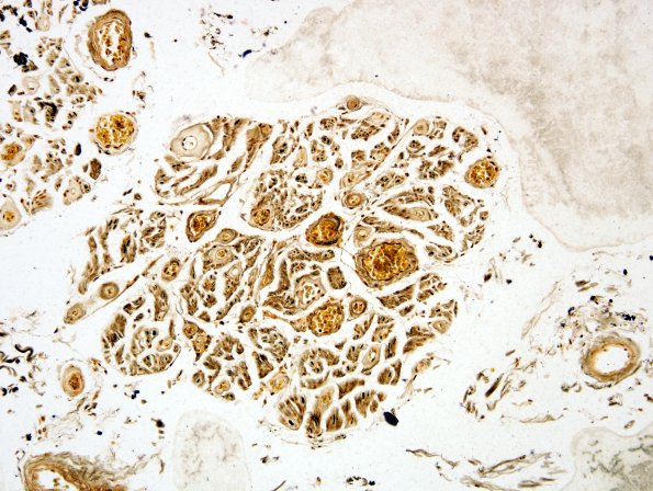
Washington University Experience | NEURODEGENERATION | ALS | 8C2 ALS (Case 8) 20x anterior root
Comparison of uninvolved dorsal root (8C1) vs depopulated ventral root (8C2). ---- Not shown: Sections of frontal and occipital neocortices show a relatively well maintained complement of neurons without concomitant astrocytic and microglial proliferation. Sections of the medulla show neuron loss and reactive gliosis in the hypoglossal nucleus. An LFB/PAS stain shows well-preserved myelination of the corticospinal tracts in the medulla. CD68 immunohistochemistry shows a diffuse increase in spinal cord microglia in all levels examined. There also is an overall increase in the number of macrophages within the white matter tracts uniformly in all sections examined. ---- Neuro Final Diagnosis: Amyotrophic lateral sclerosis, progressive muscular atrophy variant ---- Neuro Diagnosis Comment: Gross and histopathological examination of the spinal cord showed a thin caliber cord with prominent loss of lower motor neurons and reactive gliosis in the anterior horns throughout the length of the cord. Similar changes were present in the hypoglossal nucleus in sections of the medulla. There was relative preservation of the axons and myelin within the descending corticospinal tracts on examination of both routine and histochemical stained sections of the spinal cord. However, a marked increase in spinal cord microglia and a mildly selective macrophage infiltration into the lateral cortical spinal tracts was present after CD68 immunohistochemistry. Examination of the telencephalon, diencephalon, and mesencephalon were essentially unremarkable. These findings are consistent with the clinical history of amyotrophic lateral sclerosis (ALS), specifically the progressive muscular atrophy (PMA) variant, with predominantly lower motor neuron involvement relative to upper motor neuron involvement.

