Table of Contents
Washington University Experience | NEURODEGENERATION | ALS | 9A2 ALS (Case 9) H&E 1 copy
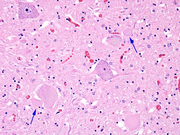
There is loss and atrophy of individual motor neurons. Noted in these two sections are large glassy axonal spheroids which often accumulate adjacent to the cell bodies (a pattern which is commonly seen in axonal dystrophy where they likely represent abnormal synapses. Rare chromatolytic neurons (arrowhead, 9A1) mimic the dystrophic axons but often have multiple branches off the cell body and peripheral marginated Nissl. Rare Bunina bodies are observed on H&E stained sections.

