Table of Contents
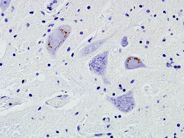
Washington University Experience | NEURODEGENERATION | ALS | 9C2 ALS (Case 9) TDP43 9
Occasional TDP43-positive skein-like and round inclusions are seen motor neurons of the anterior horns. ---- Not shown: There is minimal loss of neurons from frontal, temporal and occipital lobes, especially the precentral gyrus. No TDP43-positive inclusions are seen in the precentral gyrus. Antibodies to 'three-repeat' and 'four-repeat' tau show that modest tau pathology is composed of four-repeat tau, and only rare lesions show reactivity for three-repeat tau. No TDP43 proteinopathy or alpha-synuclein proteinopathy is seen in either the amygdala or entorhinal cortex. There is some loss of neurons in the hypoglossal nucleus. There is mild pallor of the pyramidal tracts, consistent with tract degeneration. Sparse skein-like TDP- 43-immunoreactive neuronal cytoplasmic inclusions (NCI) are identified in the motor neurons of the hypoglossal nucleus. ---- Neuro Diagnosis Comment: Consistent with the absence of cognitive deficits, the cortex of the prefrontal and temporal lobes showed no involvement. Moreover, the tau pathology in this case appears to reflect accumulation almost exclusively of four-repeat (4R) tau rather than the mixture of three-repeat (3R) and 4R Tau that is common in Alzheimer disease. The relative preservation of the substantia nigra and absence of Lewy bodies also excludes Parkinson’s disease and dementia with Lewy bodies as diagnostic considerations. The hippocampus shows marked neuronal loss and gliosis throughout the pyramidal layer of Ammon’s horn, consistent with hippocampal sclerosis. Unlike most cases of hippocampal sclerosis, no TDP43 deposits were seen in the dentate gyrus. This patient's brain also shows early-stage Alzheimer's disease-type changes, but this does not appear to have had any significant impact on cognition.

